Difference between revisions of "MGH head 1:MGH head 1"
| Line 1: | Line 1: | ||
| − | + | [[File:giorgio_phant.jpg|200px|thumb|right|]] | |
=== Citation === | === Citation === | ||
| − | + | If you use any of the material below, please don't forget to cite: | |
| − | If you use any of the material below, please cite: | ||
Nadine N. Graedel, Jonathan R. Polimeni, Bastien Guerin, Borjan Gagoski and Lawrence L. Wald (2015). "An anatomically realistic temperature phantom for radiofrequency heating measurements". Magnetic Resonance in Medicine 73(1):442–450 | Nadine N. Graedel, Jonathan R. Polimeni, Bastien Guerin, Borjan Gagoski and Lawrence L. Wald (2015). "An anatomically realistic temperature phantom for radiofrequency heating measurements". Magnetic Resonance in Medicine 73(1):442–450 | ||
| Line 13: | Line 12: | ||
The segmented head data was converted into a simplified surface model with two compartments: Brain and other soft tissues. Bone and air (both low epsilon, low conductivity) are printed together out of plastic material with similar electrical properties. | The segmented head data was converted into a simplified surface model with two compartments: Brain and other soft tissues. Bone and air (both low epsilon, low conductivity) are printed together out of plastic material with similar electrical properties. | ||
| − | === Special considerations == | + | === Special considerations === |
The sealing flange uses an o-ring. | The sealing flange uses an o-ring. | ||
[[File:Phantom_figure_CT_MR.png|400px|thumb|right|]] | [[File:Phantom_figure_CT_MR.png|400px|thumb|right|]] | ||
Revision as of 14:34, 24 March 2015
Citation
If you use any of the material below, please don't forget to cite: Nadine N. Graedel, Jonathan R. Polimeni, Bastien Guerin, Borjan Gagoski and Lawrence L. Wald (2015). "An anatomically realistic temperature phantom for radiofrequency heating measurements". Magnetic Resonance in Medicine 73(1):442–450
Download files
Click here to download the CAD model.
Phantom description
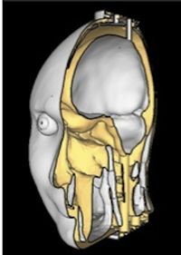
This phantom is based on a 1mm isotropic segmented head model which was labeled as described in MRI-based anatomical model of the human head for specific absorption rate mapping.
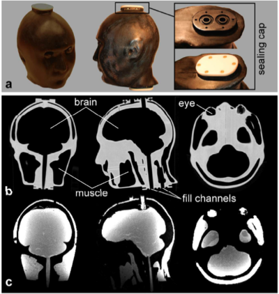
The segmented head data was converted into a simplified surface model with two compartments: Brain and other soft tissues. Bone and air (both low epsilon, low conductivity) are printed together out of plastic material with similar electrical properties.
Special considerations
The sealing flange uses an o-ring.

