DCA voxels: Difference between revisions
Jump to navigation
Jump to search
(Created page with " 400px|thumb|Segmented "Angel 001" MGH head model === Citation === None === Download files === Download the Matlab file containing the segmented vol...") |
No edit summary |
||
| (One intermediate revision by the same user not shown) | |||
| Line 1: | Line 1: | ||
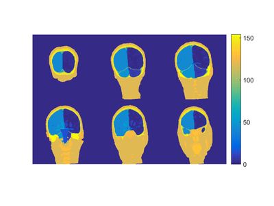
[[File: | [[File:MGH_DCA_head_model.jpg | 400px|thumb|Segmented "DCA" MGH detailed head model ]] | ||
=== Citation === | === Citation === | ||
| Line 5: | Line 5: | ||
=== Download files === | === Download files === | ||
Download the Matlab file containing the segmented volume and the tissue key [https://phantoms.martinos.org/images/ | Download the Matlab file containing the segmented volume and the tissue key [https://phantoms.martinos.org/images/7/76/MGH_DCA_head_model.zip here]. | ||
=== Phantom description === | === Phantom description === | ||
This phantom is a segmentation of a 1 mm isotropic | This head model segmentation was performed at the Center for Morphometric Analysis (CMA) at Massachusetts General Hospital (MGH). The MRI data was acquired at the Connectome scanner (3T) located at Athinoula A. Martinos Center for Biomedical Imaging. All the above work was funded by the NIH/NIBIB R21EB016449 grant. The phantom is a highly detailed segmentation of a 1 mm isotropic MRI acquisition of a healthy female into 62 tissue classes. The key between the segmented tissue classes and the tissue names is the following: | ||
* | * 0 : Unknown | ||
*White | * 2 : Left-Cerebral-White-Matter | ||
* | * 3 : Left-Cerebral-Cortex | ||
*CSF | * 4 : Left-Lateral-Ventricle | ||
* | * 5 : Left-Inf-Lat-Vent | ||
* | * 7 : Left-Cerebellum-White-Matter | ||
* 8 : Left-Cerebellum-Cortex | |||
* 10 : Left-Thalamus-Proper | |||
* 11 : Left-Caudate | |||
* 12 : Left-Putamen | |||
* 13 : Left-Pallidum | |||
* 14 : 3rd-Ventricle | |||
* 15 : 4th-Ventricle | |||
* 16 : Brain-Stem | |||
* 17 : Left-Hippocampus | |||
* 18 : Left-Amygdala | |||
* 24 : CSF | |||
* 26 : Left-Accumbens-area | |||
* 28 : Left-VentralDC | |||
* 41 : Right-Cerebral-White-Matter | |||
* 42 : Right-Cerebral-Cortex | |||
* 43 : Right-Lateral-Ventricle | |||
* 44 : Right-Inf-Lat-Vent | |||
* 46 : Right-Cerebellum-White-Matter | |||
* 47 : Right-Cerebellum-Cortex | |||
* 49 : Right-Thalamus-Proper | |||
* 50 : Right-Caudate | |||
* 51 : Right-Putamen | |||
* 52 : Right-Pallidum | |||
* 53 : Right-Hippocampus | |||
* 54 : Right-Amygdala | |||
* 58 : Right-Accumbens-area | |||
* 60 : Right-VentralDC | |||
* 62 : Right-vessel | |||
* 85 : Optic-Chiasm | |||
* 96 : Left-Amygdala-Anterior | |||
* 97 : Right-Amygdala-Anterior | |||
* 98 : Dura | |||
* 118 : Epidermis | |||
* 119 : Conn-Tissue | |||
* 120 : SC-Fat-Muscle | |||
* 122 : CSF-SA | |||
* 124 : Ear | |||
* 126 : Spinal-Cord | |||
* 127 : Soft-Tissue | |||
* 128 : Nerve | |||
* 129 : Bone | |||
* 130 : Air | |||
* 131 : Orbital-Fat | |||
* 132 : Tongue | |||
* 133 : Nasal-Structures | |||
* 135 : Teeth | |||
* 140 : Cornea | |||
* 142 : Diploe | |||
* 143 : Vitreous-Humor | |||
* 144 : Lens | |||
* 145 : Aqueous-Humor | |||
* 146 : Outer-Table | |||
* 147 : Inner-Table | |||
* 150 : R-C-S | |||
* 153 : SC-Tissue | |||
* 154 : Mastoid-Air-Cells | |||
Latest revision as of 10:05, 15 June 2016
Citation
None
Download files
Download the Matlab file containing the segmented volume and the tissue key here.
Phantom description
This head model segmentation was performed at the Center for Morphometric Analysis (CMA) at Massachusetts General Hospital (MGH). The MRI data was acquired at the Connectome scanner (3T) located at Athinoula A. Martinos Center for Biomedical Imaging. All the above work was funded by the NIH/NIBIB R21EB016449 grant. The phantom is a highly detailed segmentation of a 1 mm isotropic MRI acquisition of a healthy female into 62 tissue classes. The key between the segmented tissue classes and the tissue names is the following:
- 0 : Unknown
- 2 : Left-Cerebral-White-Matter
- 3 : Left-Cerebral-Cortex
- 4 : Left-Lateral-Ventricle
- 5 : Left-Inf-Lat-Vent
- 7 : Left-Cerebellum-White-Matter
- 8 : Left-Cerebellum-Cortex
- 10 : Left-Thalamus-Proper
- 11 : Left-Caudate
- 12 : Left-Putamen
- 13 : Left-Pallidum
- 14 : 3rd-Ventricle
- 15 : 4th-Ventricle
- 16 : Brain-Stem
- 17 : Left-Hippocampus
- 18 : Left-Amygdala
- 24 : CSF
- 26 : Left-Accumbens-area
- 28 : Left-VentralDC
- 41 : Right-Cerebral-White-Matter
- 42 : Right-Cerebral-Cortex
- 43 : Right-Lateral-Ventricle
- 44 : Right-Inf-Lat-Vent
- 46 : Right-Cerebellum-White-Matter
- 47 : Right-Cerebellum-Cortex
- 49 : Right-Thalamus-Proper
- 50 : Right-Caudate
- 51 : Right-Putamen
- 52 : Right-Pallidum
- 53 : Right-Hippocampus
- 54 : Right-Amygdala
- 58 : Right-Accumbens-area
- 60 : Right-VentralDC
- 62 : Right-vessel
- 85 : Optic-Chiasm
- 96 : Left-Amygdala-Anterior
- 97 : Right-Amygdala-Anterior
- 98 : Dura
- 118 : Epidermis
- 119 : Conn-Tissue
- 120 : SC-Fat-Muscle
- 122 : CSF-SA
- 124 : Ear
- 126 : Spinal-Cord
- 127 : Soft-Tissue
- 128 : Nerve
- 129 : Bone
- 130 : Air
- 131 : Orbital-Fat
- 132 : Tongue
- 133 : Nasal-Structures
- 135 : Teeth
- 140 : Cornea
- 142 : Diploe
- 143 : Vitreous-Humor
- 144 : Lens
- 145 : Aqueous-Humor
- 146 : Outer-Table
- 147 : Inner-Table
- 150 : R-C-S
- 153 : SC-Tissue
- 154 : Mastoid-Air-Cells
