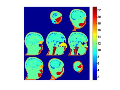
MGH head 1 voxels: Difference between revisions
Jump to navigation
Jump to search
(Created page with " 400px|thumb|Segmented "MGH head 1" head model === Citation === === Download files === Download the Matlab file containing the segmented volume and ...") |
No edit summary |
||
| Line 2: | Line 2: | ||
=== Citation === | === Citation === | ||
Makris N, Angelone L, Tulloch S, Sorg S, Kaiser J, Kennedy D, Bonmassar G. (2008), "[http://www.ncbi.nlm.nih.gov/pubmed/18985401 MRI-based anatomical model of the human head for specific absorption rate mapping]". Med Biol Eng Comput 46:1239–1251. | |||
=== Download files === | === Download files === | ||
Latest revision as of 12:16, 18 March 2016
Citation
Makris N, Angelone L, Tulloch S, Sorg S, Kaiser J, Kennedy D, Bonmassar G. (2008), "MRI-based anatomical model of the human head for specific absorption rate mapping". Med Biol Eng Comput 46:1239–1251.
Download files
Download the Matlab file containing the segmented volume and the tissue key here.
Phantom description
This phantom is a segmentation of a 2 mm isotropic MRI anatomical acquisition of a healthy male into 22 tissue classes:
- Air sinuses
- Adipose tissues
- Arteries
- Bone
- Brain
- Cerebellum
- CSF
- Ears
- Grey matter
- Head
- Humors
- Mastoids
- Nerves
- Nose
- Orbital fat
- Retina
- Skin
- Skull
- Spinal chord
- Subcutaneous fat
- Vertebral column
- White matter
